pleural effusion cat ultrasound
A chest ultrasound to look for the presence of fluid within the pleural cavity. Found with right congestive heart failure obstruction to lymphatic drainage by tissue adhesions in pleural space lung lobe torsion neoplasms and abdominal contents herniating.
Differentiating Pericardial From Pleural Effusion Animal Ultrasound Association
Four criteria have been described to differentiate ascites from pleural effusion by CT.

. Ultrasound-guided pleural effusion drainage by catheter insertion is a safe and effective procedure. The success rate is low when the effusion is loculated and septated. Accumulation of fluid in the pleural space.
This can be caused by thoracic lymphangiectasia swollen lymph vessels that leak chyle into the pleural space congestive heart failure obstruction of the cranial vena cava the major vein that returns blood to the heart from the front of the body cancer fungal infection feline heartworm. Cats presenting with pleural effusion are nearly always in respiratory distress ranging from an increased respiratory rate and effort to open mouth breathing. Global FASTÒ is the combination of AFASTÒ and its Target-organ Approach and its Abdominal Fluid Scoring System TFASTÒ for the detection of pleural and pericardial effusion pneumothorax and its 4 TFASTÒ echo views and Vet BLUEÒ the veterinary brief lung ultrasound exam a regional.
Initial treatments may vary depending on the likelihood of the specific diseases based on your pets physical examination and history. The type of pleural fluid withdrawn will enable your veterinarian to diagnose the cause of the pleural effusion. The most commonly diagnosed cause of pleural effusion in cats is chylothorax.
Pleural effusion is typically diagnosed by taking radiographs X-rays of the chest. Distinguishing Global FASTÒ from Flashing and POCUS Global FASTÒ. Ad Exclusive and Superior Vet Services from the Heart of Chelsea.
Caudal is to the left of the image. Ultrasound examination of the heart echocardiogram Laboratory tests. These four signs the diaphragm sign the displaced crus sign the interface sign and the bare area sign are reliable when only one abnormal fluid collection is present.
The cell count is 5000-300000 cellsmicroliter and contains degenerated neutrophils macrophages and bacteria. The treatment of pleural effusion ultimately will depend upon the underlying cause. Diagnostics will be necessary to confirm the cat has pleural effusion and determine a cause.
Abdominal ultrasounds were performed in 70 cats with pleural effusion and revealed concurrent abdominal effusion in 59 of these cats. Four standard effusion types recognized in addition to blood. X-ray and ultrasound imaging of the chest cavity are also very helpful in analyzing the causative factors.
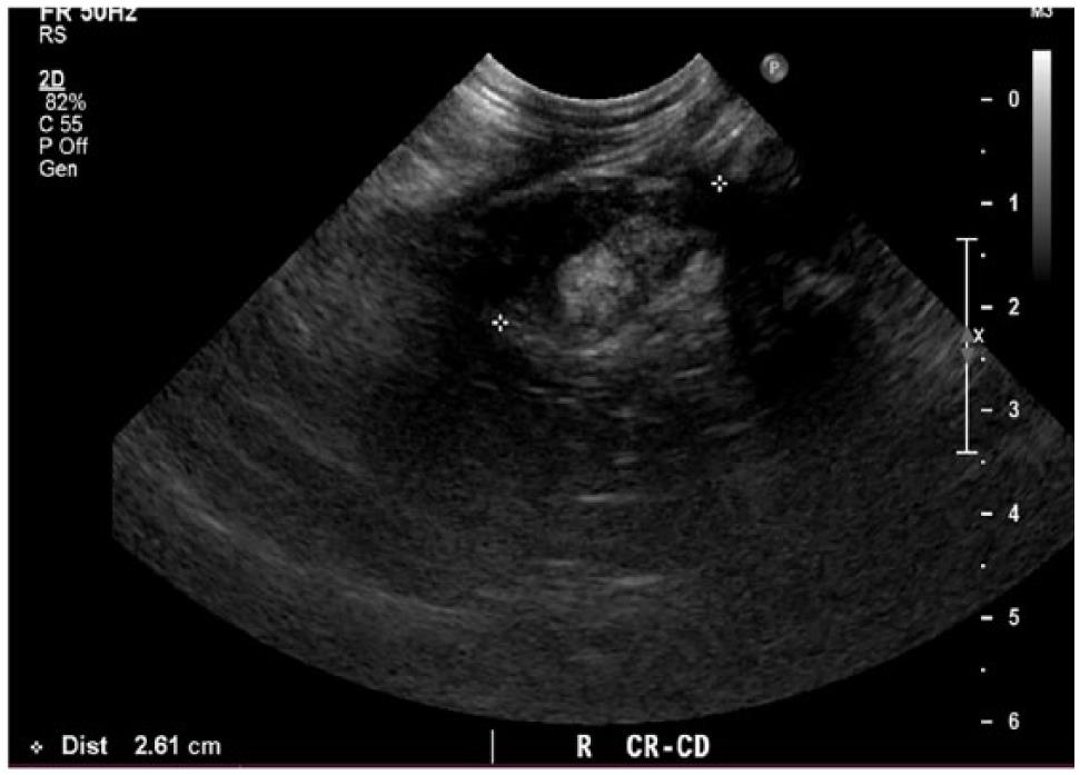
In the latter situations therapeutic intervention must be initiated quickly to prevent respiratory arrest. The caudal vena cava cvc is seen extending from the liver L to the heart H. There are a number of characteristic findings on radiographs that will help your veterinarian identify the presence of pleural effusion.
The pleural space is the gap between the two feline pleurae which line the lung and aid in breathing. Pleural effusion is present in both hemithoraces e. This non-invasive and quick test can help the veterinarian evaluate the cat quickly.
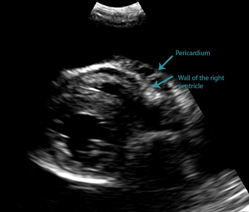
In the below clip from the Sonoscape S2 you can actually see the separation of the right ventricular free wall from the pericardium in a cat. Diverse disease processes result in sufficient fluid accumulation within the pleural space to cause respiratory compromise. In some cases ultrasound may also be.
Both the trocar and the modified Seldinger techniques can be used. The transducer is perpendicular to the ribs. Pleural effusion is an accumulation of fluid of a different nature in the pleural space of cats.
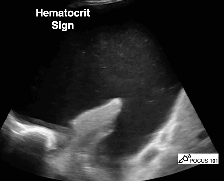
Pleural Effusion in Cats Causes Symptoms and Treatment. A cloudy or opaque yellowish-brown color the total proteins are 3-7 grdl and it contains fibrin and bacteria but not triglycerides. Focused Assessment Sonography for Trauma FAST procedure.
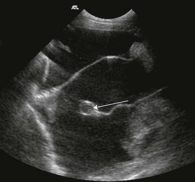
B Longitudinal ultrasound scan of the caudal thorax of a cat with pleural effusion. The color is red and opaque. Unlike with a pericardial effusion in the case of accumulation of fluid in the pleural space there is no collapse of the heart walls.
Heart of Chelsea Veterinarian Group Provides Your Pet With Superior Care. Treatment Pleural Effusion in Cats. The therapeutic intervention also provides your first diagnostic test.
Pleural effusion of septic exudate. A sample of pleural fluid obtained by piercing the cats chest cavity with a needle will be sent to the laboratory for analysis. Full Care Pet Services.
Both computed tomography CT and ultrasound US can be used to differentiate ascites from pleural effusion. Abdominal abnormalities identified on ultrasound included abdominal masses lymphadenopathy hepatic venous congestion hepatomegaly splenomegaly renal enlargement small intestinal wall thickening steatitis and pancreatitis. For this reason an abnormal accumulation of fluid in this cavity causes cats to have respiratory distress which causes them.
Determining the underlying aetiology is. The trocar technique is faster and easier. The transducer is perpendicular to the ribs.

Differentiating Pericardial From Pleural Effusion Animal Ultrasound Association

2018 Now Available For Free Download In Vetbooks Ir Vetbooks Free Veterinary Vet Veterinarian Vets Dvm Veterinary Radiology Radiology Online Textbook
Lung Ultrasound Made Easy Step By Step Guide Pocus 101

Pin Page

Acute Pleural Effusion In A Dog Clinician S Brief

Pleural Effusion And Nt Probnp In Cats

How To Ultrasound Detection Of Pleural Fluid Case Study Video Youtube

Different Types Of Pleural Effusion On Ultrasound Scan A Exudate B Download Scientific Diagram
Spontaneous Cholecystopleural Fistula Leading To Biliothorax And Sepsis In A Cat
Pleura Veterian Key
2

How To Ultrasound Detection Of Pleural Fluid Case Study Video Youtube

Pleural Effusion Ultrasound Youtube

Veterinary Echocardiography Newsletter 1 Effusions Animal Ultrasound Association

Aortic Insufficiency In Plax Color Flow Echocardiography

Pleural Effusion Empyema And Hemothorax In Three Different Patients Download Scientific Diagram

Differentiating Pericardial From Pleural Effusion Animal Ultrasound Association

Pin Page
Massive Left Sided Pleural Effusion With Floating Fibrin Deposition Download Scientific Diagram